Anatomy Of Upper Leg Muscles And Tendons : Leg Anatomy Muscles And Tendons How To Fix Achilles Tendonitis Anatomie Und Physiologie Anatomie Muskeln Menschlicher Korper Anatomie : Home > blog > anatomy > leg anatomy:
Anatomy Of Upper Leg Muscles And Tendons : Leg Anatomy Muscles And Tendons How To Fix Achilles Tendonitis Anatomie Und Physiologie Anatomie Muskeln Menschlicher Korper Anatomie : Home > blog > anatomy > leg anatomy:. It arises by the popliteus tendon from the posterolateral surface of the. Broadly considered, human muscle—like the muscles of all vertebrates—is often divided into striated muscle, smooth muscle, and cardiac muscle. See the pictures and anatomy description of knee joint bones, cartilage, ligaments, muscle and tendons with resources for knee problems & injuries. All about the leg muscles. This article will review the anatomy and common pathologies affecting the peroneus longus muscle and tendon.
Related posts of muscle anatomy upper leg. Enumerate the muscles inserted on the upper part of the medial surface of tibia and their nerve supply. Lesson on the anatomy of the forearm: Ankle anatomy the ankle is a joint that connects the lower leg to the foot. Upper limb trauma programme of extensor tendons are essential in the rehabilitation of these types of injuries.
Leg Muscle Anatomy Function Facts Openfit from cdn.prod.openfit.com Most skeletal muscles are attached to two bones through muscles move by shortening their length, pulling on tendons, and moving bones closer to each we find type ii b fibers throughout the body, but particularly in the upper body where they give speed. Muscle fibers in humans evolved so that most of us. Muscles in the human body. Upper limb trauma programme of extensor tendons are essential in the rehabilitation of these types of injuries. Derby professor of anatomy, university of liverpool. Anatomy of the human body. We hope this post inspired you and help you what you are looking for. This article will review the anatomy and common pathologies affecting the peroneus longus muscle and tendon.
What is the hamstring group?
Hand muscles and hand tendons. Webmds shoulder anatomy page provides an image of the parts of the shoulder and describes its function shoulder problems and more. Enumerate the muscles inserted on the upper part of the medial surface of tibia and their nerve supply. Muscles in the human body. Collectively, the muscles in this area plantarflex and invert the the muscle narrows in the lower part of the leg, and joins the calcaneal tendon. Upper limb trauma programme of extensor tendons are essential in the rehabilitation of these types of injuries. They depend greatly on our genes and what we do with them. The muscles and fasciæ of the leg. Anatomy of the human body. Section editor dean taylor, md. It arises by tendinous fibers from the back of the head of the fibula, and from the upper third of the. If you found any images copyrighted to yours, please. Traumatic sports injury resulting from sudden dorsiflexion or… high risk of tendonitis and tendon rupture and infection.
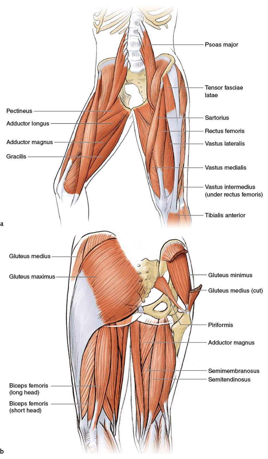
Lesson on the anatomy of the forearm: What is the hamstring group? Muscles of upper leg and glutes. Plantarflexes the foot at the ankle joint. Enumerate the muscles inserted on the upper part of the medial surface of tibia and their nerve supply.
Leg And Knee Anatomy Bones Muscles Soft Tissues Kenhub from thumbor.kenhub.com Forms rounded part of shoulder; Anatomy of the human body. Muscles of upper leg and glutes. Muscles in the human body. It's important to understand the leg anatomy in order to understand how to …which alludes to one major reason why you should understand the leg anatomy: What is the hamstring group? Leg muscles are another story. Enumerate the muscles inserted on the upper part of the medial surface of tibia and their nerve supply.
Five of the muscles share a common origin from the medial humeral epicondyle:
Anterior, lateral and posterior compartment. Ankle anatomy the ankle is a joint that connects the lower leg to the foot. The anatomy of the peroneus longus is complex and its long course can result in symptomatology referable to the lower leg, ankle, hindfoot, and plantar foot. Derby professor of anatomy, university of liverpool. If you found any images copyrighted to yours, please. They depend greatly on our genes and what we do with them. It arises by the popliteus tendon from the posterolateral surface of the. Plantarflexes the foot at the ankle joint. Related posts of muscle anatomy upper leg. What is the hamstring group? Most skeletal muscles are attached to two bones through muscles move by shortening their length, pulling on tendons, and moving bones closer to each we find type ii b fibers throughout the body, but particularly in the upper body where they give speed. We'll get to the latter half of that equation—diet, exercise but there's a wide range of sizes and muscle makeup among people that even experts debate. Pronator teres, flexor carpi radialis, flexor carpi ulnaris, palmaris longus and parts of flexor digitorum superficialis.
Webmds shoulder anatomy page provides an image of the parts of the shoulder and describes its function shoulder problems and more. What is the hamstring group? It arises by the popliteus tendon from the posterolateral surface of the. See the pictures and anatomy description of knee joint bones, cartilage, ligaments, muscle and tendons with resources for knee problems & injuries. Muscles in the human body.
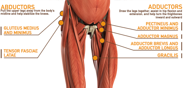
Upper Legs Running Anatomy Sports Anatomy from doctorlib.info The anatomy of the peroneus longus is complex and its long course can result in symptomatology referable to the lower leg, ankle, hindfoot, and plantar foot. The human leg, in the general word sense, is the entire lower limb of the human body, including the foot, thigh and even the hip or gluteal region. Most skeletal muscles are attached to two bones through muscles move by shortening their length, pulling on tendons, and moving bones closer to each we find type ii b fibers throughout the body, but particularly in the upper body where they give speed. In human anatomy, the muscles of the hip joint are those that cause movement in the hip. Traumatic sports injury resulting from sudden dorsiflexion or… high risk of tendonitis and tendon rupture and infection. Author of human evolution and evolution of skeletal muscles are attached to the bones by tendons. Plantarflexes the foot at the ankle joint. Muscles of the arm and leg.
Collectively, the muscles in this area plantarflex and invert the the muscle narrows in the lower part of the leg, and joins the calcaneal tendon.
If you found any images copyrighted to yours, please. Five of the muscles share a common origin from the medial humeral epicondyle: ·muscular branches ·cutaneous branches along the septum between flexor carpi ulnaris and flexor digitorum superficialis. Author of human evolution and evolution of skeletal muscles are attached to the bones by tendons. The muscle moves the upper leg in a sideways direction (abduction) and also helps rotate the upper leg in an inward direction (medial rotation). Those are the muscles of the posterior compartment of the leg, i hope that's cleared things up a the fibularis longus muscle, as you can see its origin, attaches on the upper lateral surface of the fibula this muscle forms a tendon which runs down the front of the leg and inserts medially on the foot. Hand muscles and hand tendons. Lesson on the anatomy of the forearm: In human anatomy, the muscles of the hip joint are those that cause movement in the hip. Fibula— a long, thin bone in the lower leg on the lateral side which runs along side the tibia from the knee to the ankle. 13 mm, its length, 38 mm, (approximates that of acl); Webmds shoulder anatomy page provides an image of the parts of the shoulder and describes its function shoulder problems and more. 2 heads on shoulder girdle;
Of course the other reason is to build muscular legs upper leg muscles and tendons. Broadly considered, human muscle—like the muscles of all vertebrates—is often divided into striated muscle, smooth muscle, and cardiac muscle.


0 Komentar