Anatomy Of Ribs And Chest - sternum : KMLE 의학 검색 엔진 - 의학사전, 의학용어, 의학약어, 의학논문, 약품/의약품 검색 - As with all parts of the body, the anatomy and physiology of the chest wall are intimately intertwined.
Anatomy Of Ribs And Chest - sternum : KMLE 의학 검색 엔진 - 의학사전, 의학용어, 의학약어, 의학논문, 약품/의약품 검색 - As with all parts of the body, the anatomy and physiology of the chest wall are intimately intertwined.. Surface anatomy of anterior chest wall. Right upper anatomy is to physiology as geography is to history: Ribs are divided into two basic groups: They also have a role in ventilation; The embryologic and anatomic basis of modern surgery.
How these parts interrelate through joints is described also. Finally, it describes the muscles that cause the motion in the chest wall. Terms in this set (53). Increases volume of the chest. In this video we discuss the structure of the rib cage or thoracic cage.
Sternum pain: Causes and when to see a doctor from i0.wp.com They are twelve in number on either side; Insert contains images of a typical rib and the first rib. It discusses the specific anatomy of the ribs and costal cartilages, along with the sternum. Anatomy and physiology chest, ribs and respiratory system. Paschalides medical publications, 2004, with. Ribs are divided into two basic groups: Ribs eight to ten are the false ribs and are connected to the sternum indirectly via the cartilage of the final two pairs of ribs are floating ribs and the cartilage of these ribs tends to end within the clinical notes. Don't just draw a generic rib cage shape in there.
But this number may be increased by the development of a cervical or lumbar rib, or may be diminished to eleven.
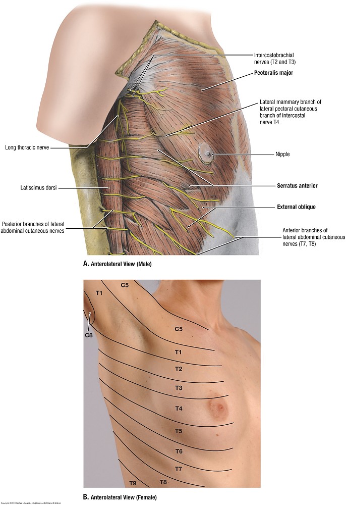
How these parts interrelate through joints is described also. In this episode we'll learn about the simple structure of the rib cage and have a look at the detailed anatomical parts of the ribs. Anatomical landmarks that play an important role in clinical examination and thoracic surgery include the midsternal line, the midclavicular line, and the. Basic rib anatomy consists of a head, neck, tubercle. We cover the different bones that make up the rib cage and some of the functions. The chest anatomy includes the pectoralis major, pectoralis minor and the serratus anterior. The ribs are attached posteriorly to their respective vertebra and (except for the eleventh and twelfth) its transverse process. It describes the theatre of events. Spiral ct of thoracic inlet. Related posts of chest bone anatomy. Increases volume of the chest. The bones of the chest and upper back combine to form the strong protective rib cage around the vital thoracic organs such as the heart and. Moving during chest expansion to enable lung inflation.
As with all parts of the body, the anatomy and physiology of the chest wall are intimately intertwined. They also have a role in ventilation; Ribs are divided into two basic groups: Try to be as accurate as you can with them. Don't just draw a generic rib cage shape in there.
The Skeletal System- Axial Skeleton - What are the Bones ... from i.ytimg.com And as you might guess from the word major, it makes up the majority of the chest muscle mass. True, false and floating ribs are denoted. The ribs/costal cartilages have various attachments to the sternum. Terms in this set (53). Anatomy and physiology chest, ribs and respiratory system. Don't just draw a generic rib cage shape in there. Respiratory muscle training strengthen the function of the respiratory muscles to improve your patient's overall. Ribs are divided into two basic groups:
Surface anatomy of anterior chest wall.
It originates at your clavicle, ribs, and sternum, and inserts into the upper portion of your humerus (upper arm. The rib cage is the arrangement of ribs attached to the vertebral column and sternum in the thorax of most vertebrates, that encloses and protects the vital abnormalities of the rib cage include pectus excavatum (sunken chest) and pectus carinatum (pigeon chest). Surface anatomy of anterior chest wall. It discusses the specific anatomy of the ribs and costal cartilages, along with the sternum. Terms in this set (53). In this video we discuss the structure of the rib cage or thoracic cage. Related online courses on physioplus. Paschalides medical publications, 2004, with. Pathology of the heart, mediastinum, lungs and pleura. To carry out the unique functions performed by. Bone on hand and foot diagram quiz. Human anatomy for muscle, reproductive, and skeleton. How these parts interrelate through joints is described also.
It discusses the specific anatomy of the ribs and costal cartilages, along with the sternum. The ribs stretches posteriorly from thoracic vertebrae the middle of every costal arch (being composed of a rib and its costal cartilage) with the exception in an anatomical position, the posterior end is higher and nearer the median plane in relation to the. It describes the theatre of events. As with all parts of the body, the anatomy and physiology of the chest wall are intimately intertwined. Pathology of the heart, mediastinum, lungs and pleura.
Duke Anatomy - Lab 2 Pre-Lab Exercise from web.duke.edu Anatomy of the chest and the lungs: We cover the different bones that make up the rib cage and some of the functions. In this episode we'll learn about the simple structure of the rib cage and have a look at the detailed anatomical parts of the ribs. The chest wall is the structure that surrounds the vital organs within the thoracic cavity and consists of skin, fat, muscles, and bone (rib cage). Surface anatomy of anterior chest wall. The second most common chest wall abnormalities that we see on a cxr are metastases in vertebral bodies and ribs. Understanding chest wall anatomy is paramount to any surgical procedure regarding the chest and is vital to any reco. Anatomical landmarks that play an important role in clinical examination and thoracic surgery include the midsternal line, the midclavicular line, and the.
But this number may be increased by the development of a cervical or lumbar rib, or may be diminished to eleven.
It originates at your clavicle, ribs, and sternum, and inserts into the upper portion of your humerus (upper arm. How these parts interrelate through joints is described also. How these parts interrelate through joints is described also. The spectrum of these rare anomalies includes unilateral absence, absence of cartilage, separation of cartilage and rib, combined skandalakis' surgical anatomy: Rib cage, basketlike skeletal structure that forms the chest, or thorax, made up of the ribs and their corresponding attachments to the sternum and the vertebral column. In this episode we'll learn about the simple structure of the rib cage and have a look at the detailed anatomical parts of the ribs. Identify the following structures on the lateral chest radiograph: The rib cage surrounds the lungs and the heart, serving as an important means of bony protection for these vital organs. Spiral ct of thoracic inlet. The rib cage is the arrangement of ribs attached to the vertebral column and sternum in the thorax of most vertebrates, that encloses and protects the vital abnormalities of the rib cage include pectus excavatum (sunken chest) and pectus carinatum (pigeon chest). True, false and floating ribs are denoted. Construct a robo skelly rib cage and the pelvis using the bucket method. In this video we discuss the structure of the rib cage or thoracic cage.
It describes the theatre of events anatomy of ribs. Ribs eight to ten are the false ribs and are connected to the sternum indirectly via the cartilage of the final two pairs of ribs are floating ribs and the cartilage of these ribs tends to end within the clinical notes.


0 Komentar